 Jeffrey Saven and Kent Blasie (Seed)
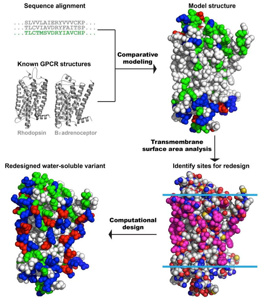
Scheme of the computational design protocol. Step 1, Comparative modeling: Starting from the sequence alignment between known GPCR structures (bovine rhodopsin and β2 adrenergic receptor) and human mu opioid receptor (MUR), a model structure of the human MUR was generated. Step 2, Identification of exposed sites in the transmembrane portion: Using the comparative model, the transmembrane lipid-exposed positions were identified (pink). Step 3, Computational design of selected exterior positions to generate a water-soluble variant: The selected exterior positions are targeted of the computational redesign with the intention of increasing the protein’s solubility and ability to be overexpressed in E. coli. Residues are colored by amino acid types: hydrophilic in green (GNQSTY); hydrophobic in white (ACFILMPVW); basic in blue (HKR); and acidic in red (DE).