Supervisor/Coordinator: Prof. Arjun Yodh
Email: yodh@physics.upenn.edu
Oversight Committee Chair: Arjun Yodh
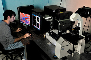 The center is a facility equipped with a wide array of optical microscopy and micro-manipulation systems to meet the needs of structural, dynamical and material characterization of soft matter, including colloids, emulsions, vesicles, liquid crystals and biomolecular materials. The facility contains:
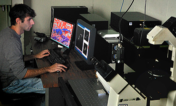
- Five optical microscopes equipped with high resolution digital video acquisition systems capable of bright field, phase contrast, high speed, and fluorescence microscopy.
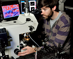
- Three high-speed confocal setups, which can be used to study three-dimensional structure of materials, and biomolecular samples.
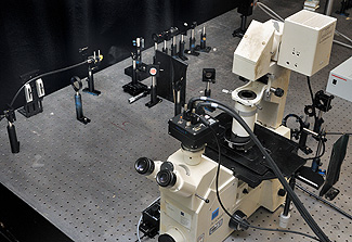
- One holographic tweezer system based on a Zeiss Axiovert 135 employs focused laser beams to manipulate microscopic objects. A spatial light modulator can generate complex patterns, and enables three-dimensional manipulation.
In addition to microscopy, the center provides other supporting optics equipment that is useful for characterization of soft materials. In particular, the center hosts a laser light scattering apparatus which is useful for angle-resolved static light scattering and photon correlation spectroscopy (i.e. quasi-elastic or dynamic light scattering spectroscopy).
EQUIPMENT:
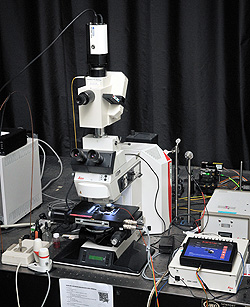
Leica DMRX
 Upright light microscope, capable of bright field, dark field, polarization, phase contrast, fluorescence, differential interference contrast (DIC) microscopy, translational stage with position reader, and objective temperature control.
- 60 fps b/w digital CCD camera (0.32 MP) (UNIQ)
- mercury lamp for fluorescence measurements
- room 333
Leica DMIR13/VT-Eye Confocal
 Inverted light microscope, capable of bright field, dark field, polarization, phase contrast, fluorescence microscopy, with objective temperature control. This microscope is attached to a VisiTech ‘VT-Eye’ confocal setup VisiTech ‘VT-Eye’
- 30 images per second (512 x 512 pixels); up to 400 images/s for reduced field of view
- Ultra-fast 3D acquisition: 256 x 256 x 100 pixel 3D image, 1 per second
- Z-scan up to 400 microns with 100 nm resolution
- Multi- wavelength excitation laser, 488, 514 and 568 nm
- VoxCell software for easy control and data management
- 60 fps b/w digital CCD camera (0.32 MP) (UNIQ)
- room 333
Zeiss Axiovert 135/Optical Tweezer
 Inverted light microscope, capable of bright field, dark field, polarization, phase contrast, fluorescence microscopy with objective temperature control. This microscope is attached to a holographic tweezer setup with spatial light modulator (SLM), capable of generating complex patterns at up to 60 frames per second with 1064 nm infrared laser
- fast (500 fps at ~1.3 MP, faster at reduced area of interest) b/w digital CMOS camera (Mikrotron)
- high-resolution (10 fps at 5 MP) b/w digital CMOS camera (EPIX)
- high-resolution (5 fps at 10 MP) b/w digital CMOS camera (EPIX)
- optical tweezers setup (Yodh group, NOT set up for general use)
- room 314
Brookhaven Instruments, BI-200SM, dynamic light scattering Capable of averaging, time-integrated intensity (classical) light scattering measurements, temperature control from 5˚C to 80˚C with stability of ±0.1 ˚C, angle selection with 0.01˚ steps
- red HeNe-laser, 15 mW
- currently not set up for static light scattering
- room 314
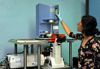
Leica DMRX A2 (Spinning Disk Confocal equipment available)
 Upright light microscope, capable of bright field, dark field, polarization, DIC, and fluorescence contrast, fully automated with motorized stage (all three dimensions), and objective temperature control.
- 60 fps b/w digital CCD camera (0.32 MP) (UNIQ)
- equipment available, but not set up:
Visitech QLC-100 Spinning Disk Confocal setup for confocal imaging with excitation wavelength of 532nm, from external CrystaLaser diode pumped crystal laser source. MetaMorph software for control and data management.
- room 312
Nikon Eclipse 200/Confocal VT Eye
 Inverted light microscope, capable of fluorescence microscopy. This microscope is attached to a VisiTech ‘VT-Eye’ confocal setup, and Bohlin Gemini rheometer (Rheology Center) VisiTech ‘VT-Eye’
- 30 images per second (512 x 512 pixels); up to 400 images/s for reduced field of view
- Ultra-fast 3D acquisition: 256 x 256 x 100 pixel 3D image, 1 per second
- Z-scan up to 100 microns with 100 nm resolution Multi- wavelength excitation laser, at 488, 569 and 633nm
- Reflection mode capability VoxCell software for easy control and data management
Light scattering facility
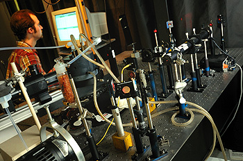 Brookhaven Instruments, BI-200SM Capable of averaging, time-integrated intensity (classical), and intensity fluctuations (Quasi-elastic) light scattering measurements, temperature control from 5 ˚C to 80 ˚C with stability of ±0.1 ˚C, angle selection with 0.01˚ steps.
Facilities users must include the following text in the acknowledgment section of their publications:
“The authors acknowledge the use of facilities supported by the Laboratory for Research on the Structure of Matter and the NSF through the University of Pennsylvania Materials Research Science and Engineering Center (MRSEC) DMR-2309043.”