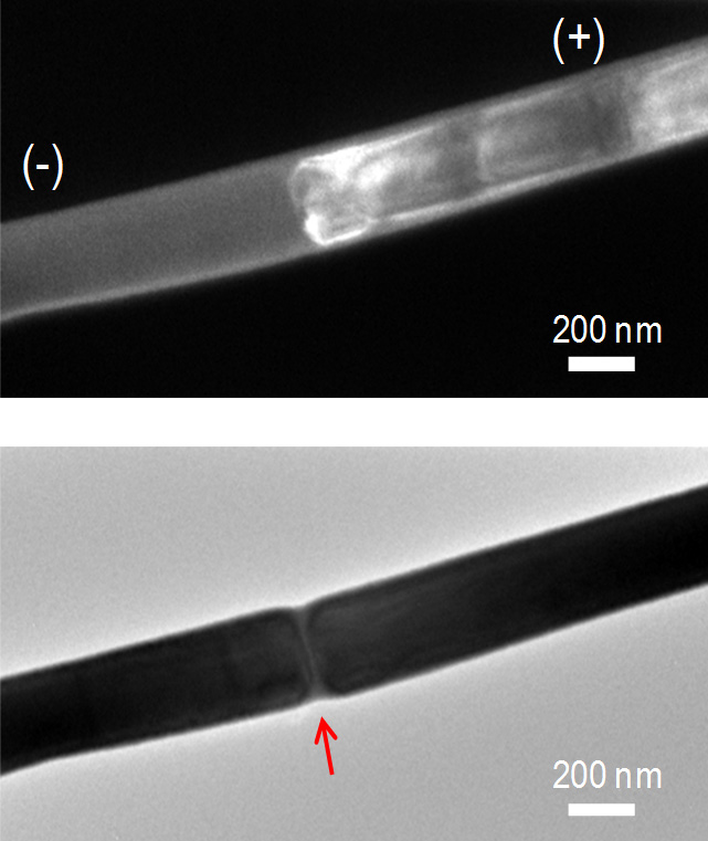
Top: Dark field transmission electron microscopy image of an electrically programmed phase change nanowire. White contrast shows the jammed dislocation cloud which has templated the nanowire along the cross-section. Bottom: Bright field image of the nanowire after amorphization. The red arrow shows the amorphous mark spanning the nanowire cross-section.