During fibrosis, collagen fibers become denser and aligned. Engineered fibrous matrices now respond to mechanical loading to densify and align fibers through inter-fiber adhesion.
Animal tissues are composed of cells attached to either the surface of a fibrous network called a basement membrane or embedded within a 3D extracellular or interstitial matrix (see Figure).
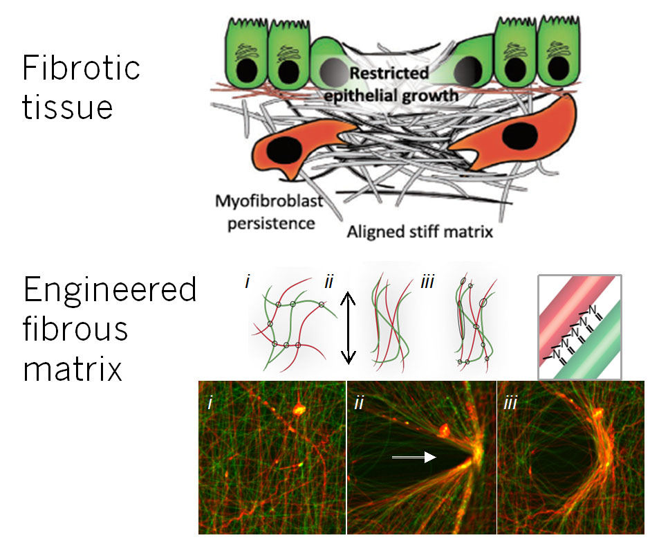
As the disease liver fibrosis progresses, the extracellular fibrous networks become denser and more aligned. These physical changes lead to different mechanical properties and structures to which cells are exquisitely sensitive.
To better understand the pathological effects of these changes during fibrosis on cells, we have engineered material platforms that mimic the extracellular matrix in tissue health and disease. As an example, we have fabricated fibrous materials that have varied mechanical properties and fiber densities when mechanically loaded due to the chemical adhesion between fibers, similar to natural extracellular matrix (see Figure).
This work exemplifies partnerships across expertise in biomaterials (Burdick), medicine (Wells), and simulation (Shenoy).