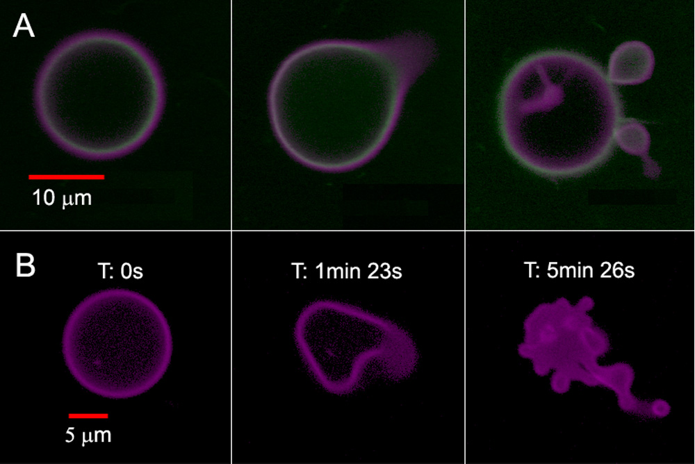
Confocal micrographs of polymersomes that membrane-disperse PZn2 (purple) and encapsulate HSAF obtained in continuous scanning mode. (A) BODIPY-FL-labeled HSAF (green, 3 mg/mL) + PZn2 vesicle, imaged using two lasers simultaneously (488 nm, 543 nm). Images proceed in time, left to right, over a period of ~5 min. (B) Unlabeled HSAF (1.5 mg/mL) + PZn2 vesicle. Vesicle imaged using three lasers simultaneously (488, 543, 633 nm). Final image of the degraded structure was not in the same plane as the original vesicle.