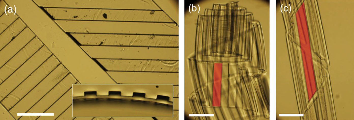
Optical microscopy images of example strips of bilayer films with thin parylene-C film deposited on the topographically patterned side of the PDMS. Scale bars: 300 mm. (a) Top-view of two dry bilayer strips with ridges at different angles. (Inset) Cross-sectional view of strip showing topographic pattern. (b, c) Swollen bilayer strips made from bilayer strips in (a) after immersion in hexadecane. The strip on the left in (a) transforms into a “tube roll” (b) after swelling. The strip on the right in (a) transforms into a “helical tube” (c) after swelling. Red shading is to guide the eye about representative ridges on the strip.